How does CEM work?
CEM uses an injection of iodinated contrast (similar to a CT exam) to highlight areas with suspicious lesions and areas of unusual blood flow using subtracted imaging.
It combines four mammography views – two low-energy and two re-combined in the same position to provide clearer visualization. The resulting image shows only where contrast has pooled, indicating areas that are potentially cancerous.
Other Benefits of Contrast-Enhanced Mammography:
- The exam takes less than seven minutes.
- It reduces the chances of false-positive exam results.
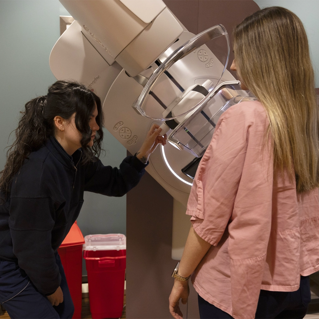
- The GE “Senographe Pristina” machine provides greater comfort and has a reduced noise level.
- Delivers personalized and optimized radiation dose based on breast density. (See our Wisely Image pledge).