Skip to content

Bone Densitometry/DXA

What is Bone Densitometry (DXA)?

Dual-energy X-ray Absorptiometry or DXA (pronounced “Dexa”) evaluates the mineral density of your bones. This typically examines the lumbar spine, hips and sometimes the forearm/wrist. Below-normal bone density (also known as “low bone mass”) can indicate loss of calcium from osteoporosis or other metabolic conditions. Although most common in women after menopause, osteoporosis sometimes affects men or younger women.

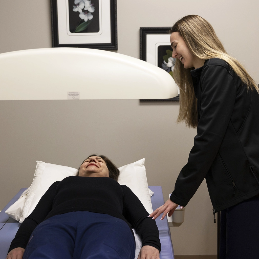
dexa bone densitometry

To detect and monitor bone mineral density, RMI offers state-of-the-art DXA to detect low bone mass (osteopenia or osteoporosis). This can also be used to monitor the effectiveness of treatments for low bone density.

In addition to scanning bone density, RMI’s radiologists have the ability to treat patients suffering from severe back pain caused by an osteoporotic fracture. A minimally invasive outpatient procedure known as vertebral augmentation ( kyphoplasty or vertebroplasty) may offer relief to patients meeting specific criteria.

Preparing for Bone Densitometry (DXA)

Unlike other medical examinations, no special preparation is required for DXA. There are no needles, dietary restrictions, or other preparation. Patients will need to bring their referring doctor’s order form, ID and insurance information, and dress in comfortable loose clothing that does not have metal.

The exam usually takes 20-30 minutes.

Resources

For more information about Cardiac CT Calcium Scores or other services offered by Regional Medical Imaging, contact us today!